
To the naked eye, a common sidewalk ant is barely a blip on the radar—a tiny, frantic dot scurrying across the pavement or trailing up a tree trunk. We crush them, brush them off our gear, and generally overlook them. But pull a single ant out of the dirt and look at it under high magnification, and that mundane insect instantly transforms into something out of a big-budget sci-fi film.
Up close, the fragile little bug vanishes. In its place stands an armor-plated creature covered in rugged, ridged skin, massive compound eyes, and heavy-duty, serrated jaws built for serious labor.
For adult outdoor enthusiasts, macro photographers, and amateur biologists, inspecting a common ant-under-microscope setup isn't just a casual observation. It’s an entirely new way to experience the natural world. Thanks to major leaps in optical tech, you no longer need a multi-thousand-dollar laboratory setup to see these details. Handheld, field-ready tools have brought high-power imaging straight to the trailhead.
The Alien Anatomy of a Household Pest
When you scale an ant up to 400x magnification, you aren't just blowing up a small image. You are revealing a highly complex, biological machine. Every single structure on an ant's body has a specific mechanical or sensory purpose.
Here is what actually comes to life when you view an ant face close up under a high-magnification lens:

-
The Mandibles (Serrated Jaws): These aren't just mouthparts; they are the ultimate multi-tool. Under a lens, you can see the distinct, saw-like teeth along the edges. Ants use these to cut through dense foliage, carry objects dozens of times their own weight, and defend the colony from rival insects.
-
Compound Eyes: An ant doesn't see the world the way we do. Their bulging eyes are actually clusters of hundreds of individual, microscopic lenses. Together, they create a wide-angle mosaic view designed primarily to detect rapid movement and shifting light patterns on the forest floor.
-
Articulated Antennae: If you watch a living ant, its antennae are constantly twitching. Under magnification, you’ll see these are highly flexible, jointed appendages. They are packed with chemical receptors, essentially acting as the insect's nose and fingertips to track pheromone trails and identify nestmates.
-
Sensory Hairs (Setae): Look closely at the legs and face, and you’ll see a dense coat of fine, translucent hairs. These aren't for warmth. They are highly sensitive tactile tools that detect subtle air currents and ground vibrations, warning the ant of approaching predators.
Why 400x Magnification is the Sweet Spot for Field Biology
There is a massive technical jump between a basic 40x jeweler's loupe and a true 400x optical inspection. Low-power magnification shows you the shape of the insect; high-power magnification shows you the story of the insect.
At 400x zoom, you begin to see things that standard macro photography lenses completely miss:
-
The deep, geometric ridges and microscopic pores scoring the ant’s chitinous exoskeleton.
-
Individual grains of flower pollen or forest dust trapped like boulders between the hairs on its legs.
-
Tiny, microscopic parasites or mites that use the ant as a personal transport vehicle.
-
The exact boundaries of the individual facets within the compound eye structure.
For anyone serious about entomology, botany, or content creation, this level of clarity changes the game. It bridges the gap between raw outdoor exploration and deep biological discovery, turning a standard hike into a deep-dive research expedition.
Redefining the Field Kit: High-Res Imaging on the Go
Historically, the biggest barrier to this level of discovery was the hardware. Traditional microscopes are heavy, fragile, and bound to a desk by a power cord. They belong in sterile labs, not in a backpack on a muddy trail.
That is where modern digital imaging completely changes the dynamic. Using a dedicated, ultra-portable microscope camera allows you to bring lab-grade optics directly into the field. Whether you are tracking a colony in your backyard or documenting specimens deep in the woods, a handheld microcam lets you inspect living specimens on site without damaging them or dragging them back to a desk.
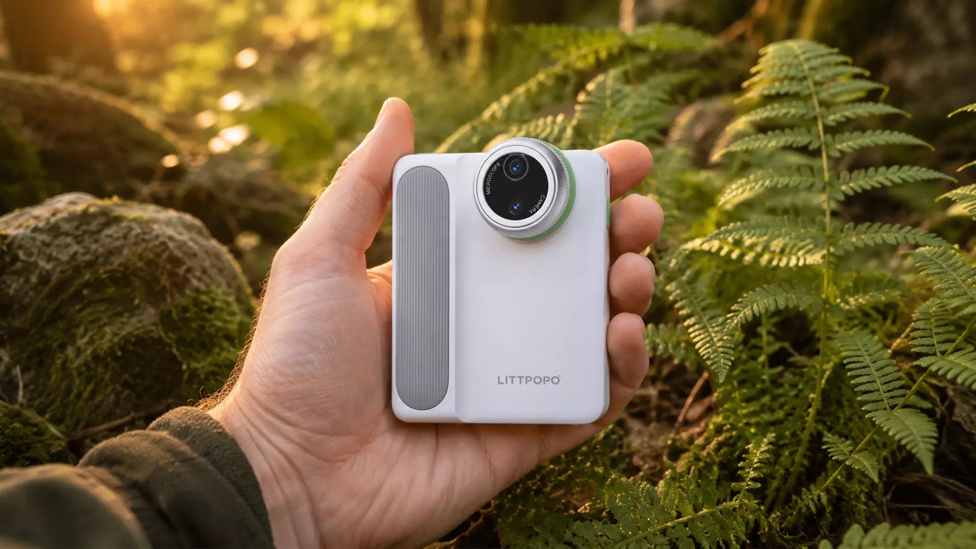
Engineered for the Rugged Explorer

High-end portable systems like the Littpopo MicroCam X1 are built specifically to bridge the gap between field mobility and professional imaging performance:
-
400X Optical Precision: Delivers the raw power needed to isolate the individual ridges on an ant's face or the microscopic details of a specimen.
-
12MP High-Resolution Stills: Freezes the chaotic motion of the micro-world into razor-sharp, publication-ready images showing every hair and texture.
-
4K Ultra HD Video: Captures living insects, moving fluids, and biological behaviors in real-time, perfect for high-end nature documentaries or social media content creation.
-
Adventure-Ready Footprint: Pocket-sized, completely wireless, and USB-rechargeable. It syncs directly to your phone or tablet, turning your mobile screen into a high-definition field monitor.
Beyond the Anthill: What Else to Track on the Trail
While an ant-under-microscope view is an incredible place to start, a portable digital microscope completely opens up your entire outdoor environment. Once you train your eye to look for the microscopic structures in nature, you'll find endless subjects on every single trail:
-
Plant Pathology: Inspect the intricate, vein-like vascular systems of leaves, or isolate early-stage fungal outbreaks and pest damage on local flora before they are visible to the naked eye.
-
Avian and Insect Mechanics: Examine the interlocking micro-hooks of a bird feather or the shimmering, shingle-like scales that give a butterfly’s wing its brilliant color.
-
Geological Formations: Look at raw minerals, sand samples, or crystalline structures inside broken rocks to see the pure composition of the local terrain.
-
Textiles and Foraged Material: Study the tight weave of technical outdoor fabrics, the spores on the underside of a wild mushroom, or the moisture droplets clinging to a moss bed.
Final Thoughts
Microscopic exploration is no longer locked behind lab doors or restricted to academics. With lightweight, high-performance tools like a portable digital microscope camera, anyone with a sense of curiosity can reveal the hidden architecture of the natural world.
The next time you see an ant moving across the trail, remember that you are looking at a masterclass in evolutionary engineering. All it takes is the right lens to step past the surface and see the extraordinary detail hidden in plain sight.